Thursday, November 21, 2013

Children that Exercise have Healthier Knees as Adults


Written by Jeff Behar & copied from: mybesthealthportal.net

Being more physically active in childhood is linked to greater knee cartilage and tibial bone area in adulthood, according to new research findings presented at the 2012 American College of Rheumatology Annual Meeting in Washington, D.C.


While physical activity in childhood is often recommended as a means to improve adult joint health and function, little evidence exists to illustrate the correlation between childhood physical performance measures and bone structure in adulthood later on. The goal of the study was to determine if physical activity in youth was associated with more knee cartilage and tibial bone area (the bone that forms the distal part of the knee joint) 25 years later, says Graeme Jones, MD, PhD, investigator in the study and professor of rheumatology and epidemiology at Menzies Research Institute in Hobart, Tasmania.
Real-time data was gathered in 1985 on the childhood physical performance in a diverse group of 298 people in Australian. Of these, 48.7 percent were female and ages ranged from 31 to 41. The participant’s knee cartilage and tibial bone area were measured using T-1 weighted, fat-suppressed magnetic resonance imaging.
Although Dr. Jones and his colleagues had questionnaire responses on the physical activity level of the children taken in 1985, they found that current-day measurements revealed more accurate information.
Adjustments were made for age, gender, body mass index (BMI) and past joint injuries that may affect the cartilage or bone area. The results showed that childhood physical activity, including physical work capacity, leg and hand muscle strength, sit-ups, and long and short runs had a significant, consistent association with greater tibial bone area. In addition, higher childhood physical work capacity measures were associated with greater tibial cartilage area. Other types of physical activity in childhood were associated with greater cartilage area, but these measures were less significant after adjusting for medial tibial bone area.
Dr. Jones and his colleagues do not know exactly why or how physical activity may build bone and cartilage years later. “The mechanism is uncertain, but I would contend that bone area gets larger to cope with the extra demands put on it by higher levels of physical activity, and then this lead to more cartilage, as cartilage covers the surface of bone,” he says.
While the study’s findings lend greater support to the effort in many developed countries to encourage children to be more physically active, cartilage and bone are still vulnerable to damage later on that could contribute to OA, says Dr. Jones.
“Physical activity is good, but if people have an injury while doing the physical activity, this is bad. So injury prevention is important. Avoiding a high body mass index is also important, and physical activity will help with this.”
This study was funded by NHMRC of Australia.

Get Strong! Stay Strong!
Chris

Saturday, November 16, 2013

Can Physical Therapy for Rotator Cuff Tears Prevent Surgery?

Copied from MikeReinold.com
Rotator cuff repair surgery and postoperative rehabilitation continue to be some of the most debated topics on the shoulder at orthopedic and physical therapy conferences.  Numerous studies have been published showing the failure rate of rotator cuff repair surgery ranges anywhere from 25-90%.
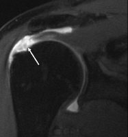
rotator cuff tearWhile this failure rate is certainly alarming, the term “failure” must be defined.  In traditional study models, success is defined as an intact rotator cuff, which makes sense.  However, one of the more interesting findings in most of these studies is that despite the “failed” repair, most patients are quite satisfied with their functional status and outcome.  This really does have to make you question how we define “failure” as patient outcomes and satisfaction seems more important than radiological findings.
These studies have sparked debate over the role of postoperative physical therapy follow rotator cuff repair surgery, with many physicians becoming more conservative and slowing down their protocols.  This obviously implies that some physicians believe that early physical therapy is the reason why failures occur.  This thinking may be flawed and factors such as tissue quality, tear severity, patient selection, surgical technique, and others may be more likely related to ultimate failure rates.
Another perspective to consider is that despite having a failed rotator cuff repair, patient satisfactions were good.  From experience, I can tell you that patients are satisfied when they:
  1. Have less pain
  2. Regain their mobility
  3. Return to functional activities
So the question really should be asked – if there is up to a 90% surgical failure rate but significant increase in satisfaction and outcomes, can physical therapy for rotator cuff tears alone without surgery be just as beneficial at helping patients reduce pain, regain mobility, and return to their activities?

Can Physical Therapy for Rotator Cuff Tears Prevent the Need for Surgery?

A recent study in the Journal of Shoulder and Elbow Surgery looked at this exact question.  The MOON Shoulder Group, which is a multi-center network of research teams around the country, followed a group of 381 patients with atraumatic full-thickness tears of the rotator cuff for a minimum of two tears.  The mean age of the patients was 62 years with a range of 31-90 years.
The patients performed 6-12 weeks of nonoperative physical therapy focusing on basic rotator cuff strengthening, soft tissue mobilization, and joint mobilizations.
At the six-week mark, patients were assessed and 9% chose to have rotator cuff repair surgery.  Patients were again assessed and the 12-week mark.  At 12-weeks, an additional 6% chose to have surgery.  In total, 26% of patients decided to have surgery by the 2-year follow-up mark.  Statistical analysis revealed that if a patient does not choose to have surgery within the first 12-weeks of nonoperative rehabilitation, they are unlikely to need to surgery.
Nearly 75% of patients avoided rotator cuff repair surgery by performing physical therapy despite having full thickness cuff tears. [Click to Tweet]
That is a pretty significant finding.

Keys to Nonoperative Rotator Cuff Rehabilitation

The results of this study could have a large impact on how we treat rotator cuff tears.  Physical therapy should be attempted prior to surgery, even in the case of a full thickness tear.  To maxmize these outcomes, a comprehensive rehabilitation program should be developed.  When working on patients with rotator cuff tears, I tend to focus on 3 key areas.

Restore Shoulder Mobility

This includes both passive and active mobility.  For passive mobility, it seems to me that shoulder range of motion is gradually lost as the rotator cuff symptoms increase.   Perhaps it is a pain avoidance strategy, disuse, or some other factor.  You’ll often find glenohumeral joint capsule hypomobility and soft tissue restrictions.  Soft tissue mobilization, joint mobilizations, and range of motion exercises should be designed based on the specific loss of motion exhibited by the patient.

Restore The Ability of the Rotator Cuff to Dynamically Stabilize

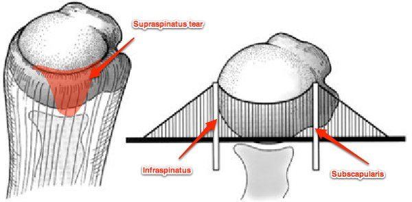
This is essentially the same as restoring active mobility of the shoulder.  The rotator cuff has to function properly to allow active mobility without restrictions.  In a previous article, I discussed the suspension bridge concept and how you can have a rotator cuff tear without symptoms.  You can see in this diagram that if you have properly functioning anterior and posterior rotator cuff muscles, you can often still elevate the arm despite a tear to the supraspinatus.
rotator cuff suspension bridge concept
Exercises designed to enhance strength and dynamic stability of the shoulder should be incorporated.  In my experience external rotation strength tends to be the most limited and needs to most attention.

Reduce the Impact of the Kinetic Chain

In addition to restore mobility and stability of the shoulder, you should also consider the impact of the kinetic chain on shoulder function.  Read my past article on the different types of shoulder impingement to understand some of these concepts.  Any dysfunctions of the scapulothoracic joint, cervical spine, thoracic spine, and lumbopelvic complex should be assessed.  These areas all have a significant impact on the alignment, mobility, and stability of the glenohumeral joint.
If you want to learn more about how I perform nonoperative rehabilitation for rotator cuff tears, I have a past webinar on shoulder impingement that discusses many of the same keys to treatment.
Using these principles, you can formulate a rehabilitation program that could potentially save 75% of people with rotator cuff tears from needed rotator cuff repair surgery.  Hopefully studies like this will continue to shed light on the impact physical therapy can have on the satisfaction and outcomes of patients with rotator cuff tears, with or without surgery.

Mikes website is chalk full of great info!
Get Strong! Stay Strong!
Chris

Monday, November 11, 2013

Essential Amino Acids and Knee Replacement


Copied from:   J Clin Inves 2013

Background. By the year 2030, 3.48 million older U.S. adults are projected to undergo total knee arthroplasty (TKA). Following this surgery, considerable muscle atrophy occurs, resulting in decreased strength and impaired functional mobility. Essential amino acids (EAAs) have been shown to attenuate muscle loss during periods of reduced activity and may be beneficial for TKA patients.

Methods. We used a double-blind, placebo-controlled, randomized clinical trial with 28 older adults undergoing TKA. Patients were randomized to ingest either 20 g of EAAs (n = 16) or placebo (n = 12) twice daily between meals for 1 week before and 2 weeks after TKA. At baseline, 2 weeks, and 6 weeks after TKA, an MRI was performed to determine mid-thigh muscle and adipose tissue volume. Muscle strength and functional mobility were also measured at these times.

Results. TKA patients receiving placebo exhibited greater quadriceps muscle atrophy, with a –14.3 ± 3.6% change from baseline to 2 weeks after surgery compared with –3.4 ± 3.1% for the EAA group (F = 5.16, P = 0.036) and a –18.4 ± 2.3% change from baseline to 6 weeks after surgery for placebo versus –6.2 ± 2.2% for the EAA group (F= 14.14, P = 0.001). EAAs also attenuated atrophy in the nonoperated quadriceps and in the hamstring and adductor muscles of both extremities. The EAA group performed better at 2 and 6 weeks after surgery on functional mobility tests (all P < 0.05). Change in quadriceps muscle atrophy was significantly associated with change in functional mobility (F = 5.78, P = 0.021).

Conclusion. EAA treatment attenuated muscle atrophy and accelerated the return of functional mobility in older adults following TKA.

Love to see medicine and nutrition come together!

Get Strong! Stay Strong!